The West African Microscopy and Bio-Imaging Analysis Network (WAMBIAN) has ended a two-week training course in bio-imaging for research scientists at the West African Centre for Cell Biology of Infectious Pathogens (WACCBIP). The course which started on September 26, 2022, brought together 19 participants from countries across West Africa, including Benin, Togo, Nigeria and Ghana, to train them on microscopy techniques and expose them to various approaches to bio-imaging and bio-image analysis.
The course facilitators were Dr Yaw Aniweh, Head of Technology at WACCBIP; Dr Peran Hayes from the Berlin Institute for Medical Systems Biology; Dr Elena Remacha who previously worked for Leica Microsystems and Dr Alassane Mbengue, a Research Associate from Institut Pasteur Dakar, Senegal.
Some of the topics covered during the training included optics and basic microscopy, fluorescence microscopy and super-resolution imaging techniques, imaging biosamples, and basic image analysis with Fiji ImageJ. Dr Peran engaged participants on the fundamental features of light in terms of ray, wave, and particle optics, focusing on image formation using lenses. He also enlightened the participants on Point Spread Functions (PSFs), resolution limits and approaches to super-resolution microscopy.
For her part, Dr Elena took the participants through the concept of confocal microscopy explaining how by use of a laser, scan mirrors, a pinhole and PMT detector optical sectioning can be achieved. She also spoke on objective lens errors, including chromatic and spherical aberrations among other issues often faced by microscopists. She also highlighted the need to think through the selection of lens immersion medium and coverslip thickness as a way to effectively mitigate spherical aberration. The participants were also introduced to the photoelectric effect, Nyquist's criteria for good sampling and various contrast techniques used for visualization. These included bright-field, phase contrast, polarization contrast, and differential interference contrast (DIC) microscopy, as well as darkfield and fluorescence techniques.

Figure 1: Dr Elena Remacha presenting on visualization topics and troubleshooting issues.
Dr Alassane delivered a presentation on the relationship between microscopy and lab work to enhance the reliability of experimental results. He discussed how studying molecular dynamics through microscopy is possible via the use of modified fluorescence visualization techniques such as Fluorescence Recovery after Photobleaching (FRAP), Fluorescence Resonance Energy Transfer (FRET), and Bimolecular Fluorescence Complementation (BiFC), among others.
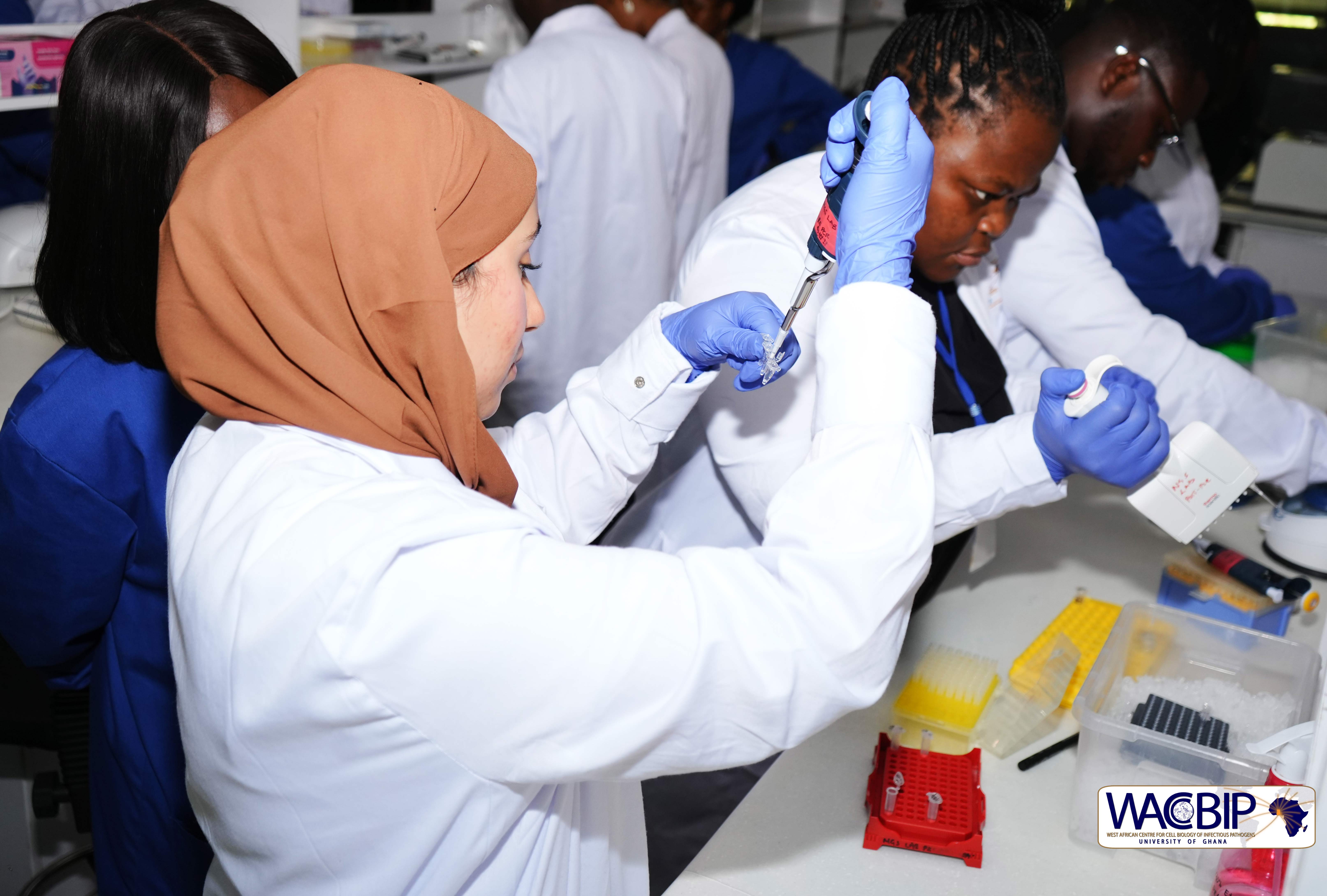
During some of the many practical sessions of the workshop, participants gained hands-on experience in working with malaria parasite labelling in infected red blood cells. The participants were given hands-on microscope assembly with 3D printable open flexure microscopes (they worked in groups in building 5 of these microscopes). The labelled malaria parasites were imaged using the institute’s Zeiss LSM 800 confocal and their own self-built microscopes.
The second week of the workshop focused on bio-image analysis. Mixing theoretical and practical sessions, participants were taken through key concepts in digital images, and methods to extract quantitative information to address biological questions.

Figure 2: Dr Peran guiding participants through visualization with the confocal microscope.

Figure 3: Leishmania parasites stained and visualized with fluorescence microscopy.
In small groups, they were then set different challenges to design automated approaches to quantify features of the images they had taken. On the final day, each group presented their solutions to the class, initiating an active and engaging discussion. Dr Peran also gave a talk on research in Germany and funding opportunities available to allow African students to come to Germany to enhance their research careers. This presentation was done in the context of the MDC Back to the Roots programme that supported the workshop.

Figure 4: Workshop participants assembling a 3D printed microscope with Dr. Elena

Figure 5: Workshop participants assembling a 3D printed microscope with Dr. Peran
At the end of the two weeks, short interview sessions were held with the participants to get feedback on workshop activities. Dr Murtala Bindawa Isah, a post-doctoral fellow at the Biomedical Science Research and Training Centre, Yobe State-University of Damaturu, Nigeria, expressed his satisfaction with the entire programme; "The content of the workshop is excellent for starters on microscopy. Additionally, it has been designed to be easily modified for more advanced participants. I would highly recommend the workshop for microscopy enthusiasts.”
Another participant, Philip Ilani, PhD Fellow at the West African Centre for Cell Biology of Infectious Pathogens (WACCBIP), said, "It was an exciting and knowledge-packed two-week microscopy and image analysis workshop. I learnt a lot and would recommend the workshop to continue yearly.”
The workshop was supported by WACCBIP, MDC-Berlin; Back to the Roots – Opportunities for studying in Germany, TReND in Africa, AHF Analysentechnik and The Open Flexure project.


